Ischial tuberosity
| Ischial tuberosity | |
|---|---|
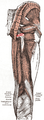
 Capsule of hip-joint (distended). Posterior aspect. (Ischial tuberosity visible at bottom left.) | |
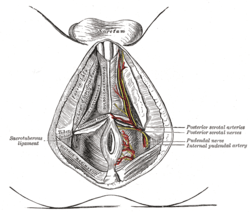 The superficial branches of the internal pudendal artery. (Ischial tuberosity visible at center left.) | |
| Details | |
| Identifiers | |
| Latin | Tuber ischiadicum, tuberositas ischiadica |
| TA | A02.5.01.204 |
| FMA | 17010 |
The ischial tuberosity (or tuberosity of the ischium, tuber ischiadicum), also known informally as the sit bones, or as a pair the sitting bones[1] is a large swelling posteriorly on the superior ramus of the ischium. It marks the lateral boundary of the pelvic outlet.
When sitting, the weight is frequently placed upon the ischial tuberosity.[2] The gluteus maximus provides cover in the upright posture, but leaves it free in the seated position.[3]
Divisions
The tuberosity is divided into two portions: a lower, rough, somewhat triangular part, and an upper, smooth, quadrilateral portion.
- The lower portion is subdivided by a prominent longitudinal ridge, passing from base to apex, into two parts:
- The outer gives attachment to the adductor magnus
- The inner to the sacrotuberous ligament
- The upper portion is subdivided into two areas by an oblique ridge, which runs downward and outward:
- From the upper and outer area the semimembranosus arises
- From the lower and inner, the long head of the biceps femoris and the semitendinosus
Additional images
 Muscles of the gluteal and posterior femoral regions, with ischial tuberosity highlighted in red.
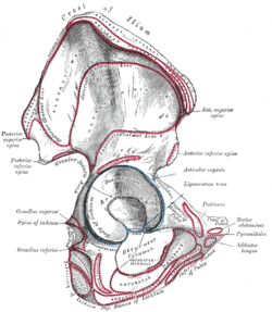 Right hip bone. External surface.
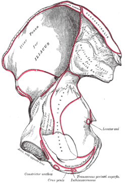 Right hip bone. Internal surface.
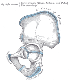 Plan of ossification of the hip bone.
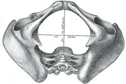 Diameters of inferior aperture of lesser pelvis (female).
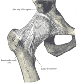 Right hip-joint from the front.
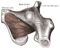 The Obturator externus.
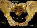 Anterior view of the pelvis with the ischial tuberosity labelled in the lower part of the image
See also
Notes
- ↑ Sills, Franklyn (2004). Craniosacral Biodynamics: The Primal Midline and the Organization of the Body (revised, illustrated ed.). Berkeley, CA: North Atlantic Books. p. 99. ISBN 1-55643-390-5.
- ↑ Goossens (2005), pp 895–982
- ↑ Platzer (2004), p 236
References
This article incorporates text in the public domain from the 20th edition of Gray's Anatomy (1918)
- Goossens R, Teeuw R, Snijders C (2005). "Sensitivity for pressure difference on the ischial tuberosity". Ergonomics. 48 (7): 895–902. doi:10.1080/00140130500123647. PMID 16076744.
- Platzer, Werner (2004). Color Atlas of Human Anatomy, Vol. 1: Locomotor System (5th ed.). Thieme. ISBN 3-13-533305-1.
External links
- Anatomy photo:41:st-0204 at the SUNY Downstate Medical Center - "The Female Perineum: Bones"
- Anatomy photo:17:os-0114 at the SUNY Downstate Medical Center - "Major Joints of the Lower Extremity: Hip bone (lateral view)"
- pelvis at The Anatomy Lesson by Wesley Norman (Georgetown University) (pelvisposterior, pelvislateral, pelvisinside)
This article is issued from Wikipedia - version of the 11/29/2016. The text is available under the Creative Commons Attribution/Share Alike but additional terms may apply for the media files.